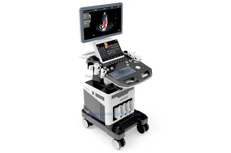
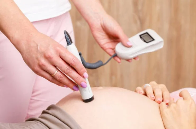
3D/4D High-End Ultrasound
3D/4D ultrasound provides detailed images of the body in real time. It is especially helpful for monitoring pregnancies, allowing parents to see their baby’s movements and features clearly.
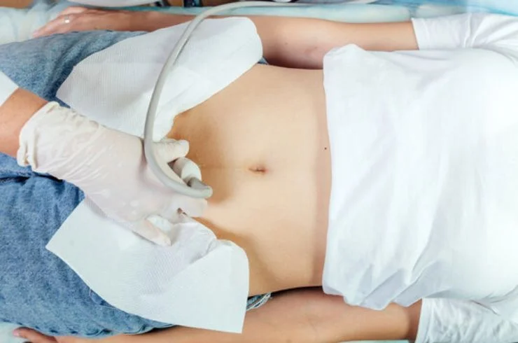
USG Abdomen
An abdominal ultrasound helps examine organs like the liver, kidneys, gallbladder, and pancreas. It’s a non-invasive, painless way to detect conditions such as tumors, gallstones, or infections.

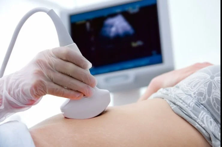
3D/4D Fetal Anomaly Scan (Level II Scan)
This detailed scan is performed during pregnancy to check for any physical abnormalities in the fetus. It provides a comprehensive view of fetal development and health.
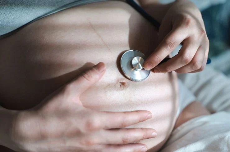
Fetal Well-Being
A fetal well-being scan assesses the baby's health inside the womb, checking for proper growth, movement, and amniotic fluid levels, ensuring everything is progressing as expected.
Fetal Well-Being with Colour Doppler
This scan uses Doppler technology to assess blood flow to the fetus, ensuring that the baby is receiving enough oxygen and nutrients for healthy development throughout pregnancy.
NT Scan (Level I Scan)
The NT (Nuchal Translucency) scan is conducted in early pregnancy to assess the risk of genetic conditions like Down syndrome. It measures the fluid at the back of the baby’s neck.
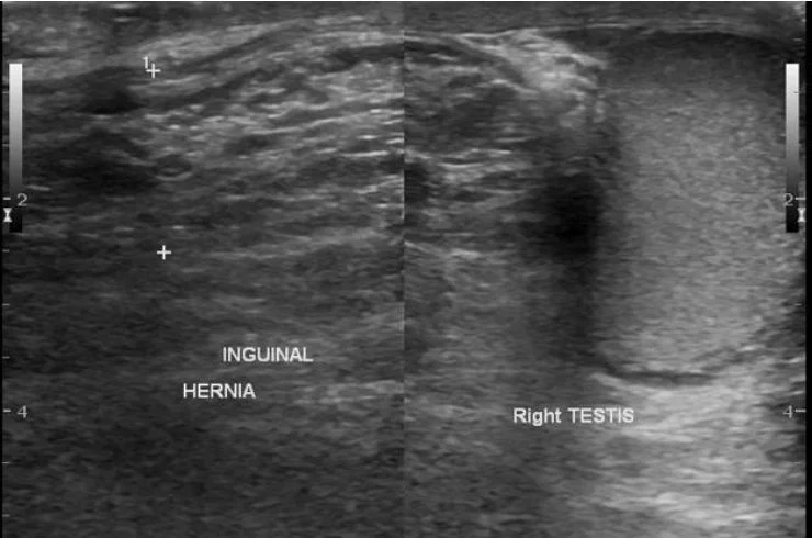
HR-USG Inguino-Scrotum
A high-resolution ultrasound of the inguino-scrotal area helps detect abnormalities in the groin and scrotum, such as hernias or testicular issues, providing clear and detailed images for diagnosis.
HR-USG Soft Tissue Lump/Swelling
This high-resolution ultrasound is used to evaluate lumps or swelling in soft tissues, helping identify the cause, such as infections, cysts, or tumors, in a non-invasive manner.
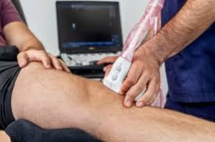
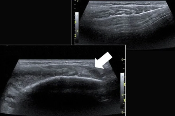
HR-USG Musculoskeletal (Joints)
A high-resolution musculoskeletal ultrasound evaluates joint conditions, such as arthritis, tendon injuries, or ligament tears, offering a precise view of the affected area for accurate treatment.
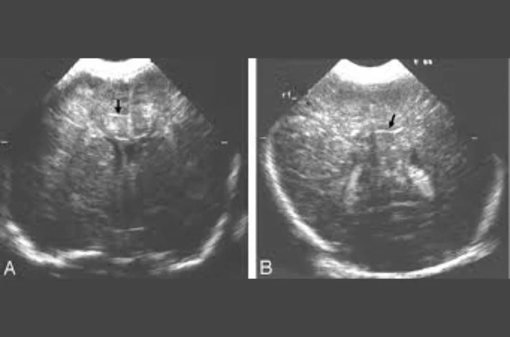
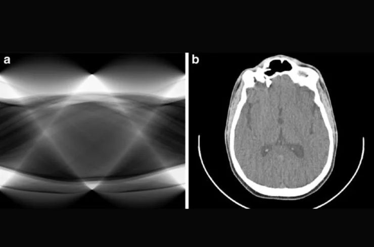
Cranial Sonography (Infant)
Cranial ultrasound is used in infants to assess brain structures, particularly in premature babies, to detect abnormalities like hemorrhages, hydrocephalus, or other developmental issues.
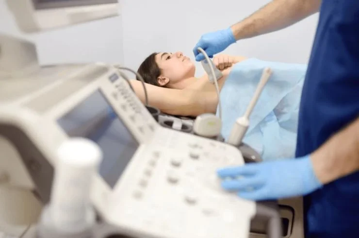
HR-USG Breast
A high-resolution breast ultrasound helps detect abnormalities like lumps, cysts, or tumors in the breast tissue, offering a non-invasive way to assist in early breast cancer detection.
HR-USG Neck/Parotid Gland with Doppler
This ultrasound assesses the neck and parotid glands for abnormalities, using Doppler to check blood flow in these areas, detecting conditions like tumors, infections, or blockages.
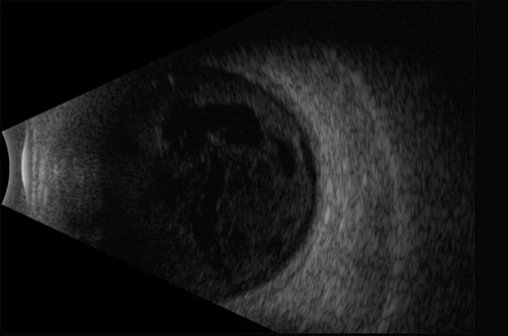
HR-USG Orbit/Eye
High-resolution ultrasound of the orbit/eye helps diagnose eye-related issues such as tumors, retinal detachments, or other abnormalities in the eye structure, offering detailed imaging.
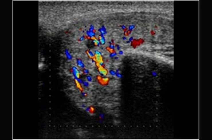
Colour Doppler Carotid Artery
A Doppler scan of the carotid arteries assesses blood flow to the brain, detecting blockages or narrowing, which can help prevent strokes and other cerebrovascular conditions.
Colour Doppler Peripheral Vessels
This scan evaluates the blood flow in peripheral vessels, such as the arms and legs, to detect blockages, clots, or poor circulation, helping diagnose vascular conditions.
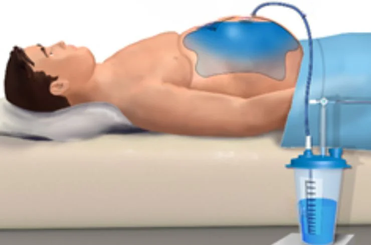
USG Guided Pleural/Ascitic Taping/FNAC
This ultrasound-guided procedure helps drain fluid from areas like the chest (pleural) or abdomen (ascites) and assists in performing Fine Needle Aspiration Cytology (FNAC) for diagnosis.

High-Frequency Digital X-ray
Digital X-rays use high-frequency radiation to capture detailed images of bones and tissues. They are used to diagnose fractures, infections, or other internal issues quickly and accurately.

Barium Meal X-ray
A barium meal X-ray examines the digestive tract by using a contrast material (barium) to highlight the esophagus, stomach, and intestines, helping detect ulcers, tumors, or blockages.
Sinogram
A sinogram is an imaging test used to investigate abnormal openings or sinuses in the body, such as fistulas, by injecting a contrast dye and capturing X-ray images of the area.
Fistulogram
This procedure evaluates fistulas (abnormal connections between organs) using a contrast dye to visualize the pathway of the fistula on an X-ray, aiding in diagnosis and treatment planning.
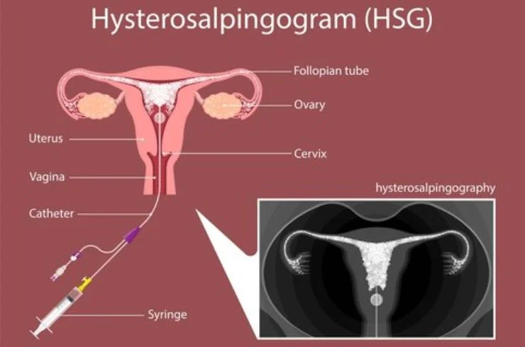
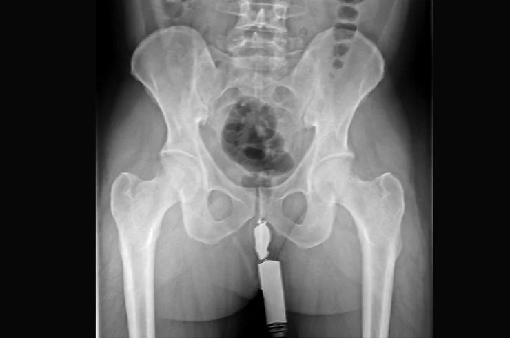
HSG
HSG (Hysterosalpingography) is a specialized X-ray procedure that examines the uterus and fallopian tubes to assess fertility issues, helping detect blockages, abnormalities, or structural problems.
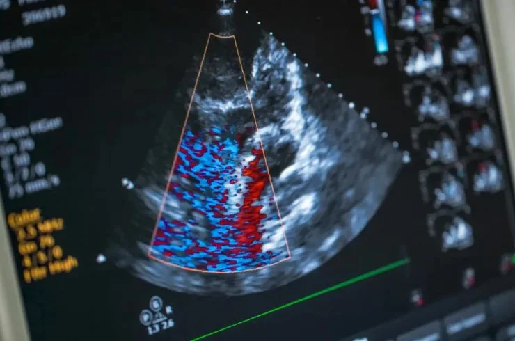
Echocardiography
Echocardiography uses ultrasound to create images of the heart, allowing doctors to assess heart function, detect abnormalities, and diagnose conditions like heart disease or valve issues.
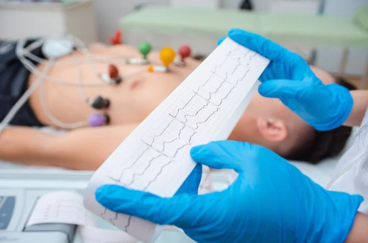
ECG (Electrocardiogram)
An ECG records the heart's electrical activity, helping detect abnormal rhythms, heart attacks, or other cardiac issues by monitoring the heart’s beats and electrical impulses.